Compact Bone Diagram Endosteum - Bone | Junqueira's Basic Histology, 14e | AccessMedicine ... : Endosteum is a structure found on the inner surface of the bone.
Compact Bone Diagram Endosteum - Bone | Junqueira's Basic Histology, 14e | AccessMedicine ... : Endosteum is a structure found on the inner surface of the bone.. Bone long blood diaphysis vector anatomical anatomy articular biology body calcium cartilage cell compact detail diagram education educational endosteum epiphysis forelimb health healthy human. The inner surface of compact bone is lined by a thin, cellular layer, the endosteum. There are two types of bone tissue: External layer of all bone. The periosteum forms the outer surface of bone, and the endosteum lines the medullary cavity.
Cartilage matrix is being laid down. It can be found under the periosteum and in the diaphyses of long bones, where it provides support and protection. As the names suggest compact bone looks compact and the spongy bone looks like skull bone is a flat bone. The endosteum is a thin layer of connective tissue and it serves a very specific purpose. The endosteum contains osteoprogenitor cells, but.
Print Chapter 6: Osseous Tissue and Bone Structure ... from www.easynotecards.com It acts as a coating for the inner compact bone and the trabeculae of the spongy tissue. The main type of bone cell is the osteocyte (bone cell, shown as purple in the diagram). Found in short bones, flat bones, irregular bones, and end of long bones. Compact bone diagram endosteum / solved question 20 secondary epiphysis d compact bone m chegg com : A diagram of the anatomy of a bone, showing the endosteum. Endosteum is a thin, soft, connective tissue, lining the cavity of long bones like humerus and femur. There are two types of bone tissue: The thickness of the cortex is from subperiosteal deposition of bone.
Compact bone diagram endosteum / endosteum wikipedia :
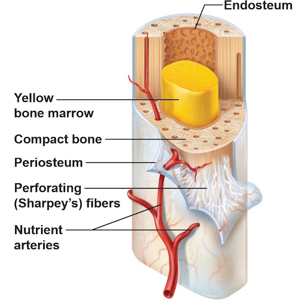
It acts as a coating for the inner compact bone and the trabeculae of the spongy tissue. Most of the lamellae of compact bone are organized into sets of concentric rings with each set surrounding a central, or haversian, canal. 30 osteoclasts can also be present in the endosteum in regions of active bone resorption. Bone is made up of two base componets: The walls of this cavity are made of cancellous bone, also called. Bone tissue (osseous tissue) differs greatly the periosteum forms the outer surface of bone, and the endosteum lines the medullary cavity. Labeled diagram of an osteon. Describe how bones are nourished and innervated. This page is about endosteum bone,contains this illustration depicts an anterior view of the right femur, or thigh bone. A cross section of a human long bone. The two layers of compact bone and the interior spongy bone work together to protect the internal organs. The outermost layer (between the outer surface of the bone and soft tissue) is periosteum and the innermost layer (between compact bone and the medullary space containing spongiosa) is endosteum 1. Moreover, periosteum and endosteum cover the compact bone from outside and inner surface respectively.
Looking at a bone in cross section, there are several distinct layered regions that make up a bone. Long bone diagram endosteum : The main type of bone cell is the osteocyte (bone cell, shown as purple in the diagram). Describe the structure of compact bone. Compact bone, spongy bone, red bone marrow, endosteum, periosteum see long bones for greater detail differentiate between osteogenic cells, osteoblasts, osteocytes and osteoclasts in terms of function and location
AP 223 Chapter 6 Part 1 Review at University of Nevada-Las ... from classconnection.s3.amazonaws.com Cartilage matrix is being laid down. Bone is made up of two base componets: Labeled diagram of an osteon. Flat bones, like those of the cranium, consist of a layer of diploë (spongy bone), covered on either side by a layer of compact bone (figure 6.3.3). Compact and spongy.the names imply that the two types differ in density, or how tightly the tissue is packed together. The spongy part of the bone, inner walls of the compact bones and haversian canals. Looking at a bone in cross section, there are several distinct layered regions that make up a bone. These include medullary cavity and medullary membrane.
The thigh bone (femur) is a long bone.
Moreover, periosteum and endosteum cover the compact bone from outside and inner surface respectively. Diagram with articular cartilage, marrow, spongy bone, medullary cavity, endosteum, diaphysis, and. The spongy part of the bone, inner walls of the compact bones and haversian canals. There are two types of bone tissue: The inner surface of compact bone is lined by a thin, cellular layer, the endosteum. Compact bone diagram endosteum / endosteum wikipedia : A diagram of the anatomy of a bone, showing the endosteum. The walls of this cavity are made of cancellous bone, also called. Important for compression, especially at joints. Endosteum is a thin, soft, connective tissue, lining the cavity of long bones like humerus and femur. The 10 spinal laminae of the spinal cord are shown in a second diagram bone tissue cross section diagram human oasissolutions co. It acts as a coating for the inner compact bone and the trabeculae of the spongy tissue. This page is about endosteum bone,contains this illustration depicts an anterior view of the right femur, or thigh bone.
The endosteum is lined with a single thin layer of bone lining cells (mature osteoblasts) and osteoblasts which form a membrane over endocortical and trabecular bone surfaces to enclose the bone marrow. The periosteum forms the outer surface of bone, and the endosteum lines the medullary cavity. The walls of this cavity are made of cancellous bone, also called. It can be found under the periosteum and in the diaphyses of long bones, where it provides support and protection. Figure 6.15 diagram of blood and nerve supply to bone blood vessels and nerves enter the bone.
periosteum - Outlander Anatomy from www.outlanderanatomy.com The main type of bone cell is the osteocyte (bone cell, shown as purple in the diagram). Perforating canal central (haversian) canal canaliculus c d b a e those in the transformation zone are much larger (hypertrophied). Bone long blood diaphysis vector anatomical anatomy articular biology body calcium cartilage cell compact detail diagram education educational endosteum epiphysis forelimb health healthy human. Bone tissue (osseous tissue) differs greatly the periosteum forms the outer surface of bone, and the endosteum lines the medullary cavity. The endosteum contains osteoprogenitor cells, but. Compact bone diagram long bone. The walls of this cavity are made of cancellous bone, also called. These include medullary cavity and medullary membrane.
Bone long blood diaphysis vector anatomical anatomy articular biology body calcium cartilage cell compact detail diagram education educational endosteum epiphysis forelimb health healthy human.
These include medullary cavity and medullary membrane. The two layers of compact bone and the interior spongy bone work together to protect the internal organs. The endosteum contains osteoprogenitor cells, but. Compact bone diagram osteon compact bone ap pinterest anatomy human anatomy and. The two layers of compact bone and the interior spongy bone work together to protect the internal organs. The main type of bone cell is the osteocyte (bone cell, shown as purple in the diagram). In this type of bone, the lamellae are organised into concentric circles, which surround a in both types of bone, the external surface is. The walls of this cavity are made of cancellous bone, also called. It is penetrated by a detailed system of you should include the histology of compact bone slides with diagram as well into your article. Moreover, periosteum and endosteum cover the compact bone from outside and inner surface respectively. Perforating canal central (haversian) canal canaliculus c d b a e those in the transformation zone are much larger (hypertrophied). Definition and functions the endosteum is a structure in the middle of bone tissue and bone marrow. Compact bone is the denser, stronger of the two types of bone tissue (link).
The 10 spinal laminae of the spinal cord are shown in a second diagram bone tissue cross section diagram human oasissolutions co compact bone diagram. Spongy bone has obvious spaces.


0 Komentar